Foot Muscles Mri : Abductor hallucis muscle | Radiology Reference Article | Radiopaedia.org. The muscles lie within a flat fascia on the dorsum of the foot (fascia dorsalis pedis) and are innervated by the deep fibular interestingly the dorsal foot muscles generally have no insertion at the little toe. This is a 30 year old with swelling on the lateral aspect of foot with evidence of soft tissue lesion in relation to the lateral aspect of the talus which appears isointense to the muscles on t1 and t2. Subscribe to foot & ankle problems. Musculoskeletal system | muscle structure and function. Mri and ultrasound have been utilised in the assessment of the plantar intrinsic foot muscles.
The muscles acting on the foot can be divided into two distinct groups; These muscles begin and attach within the skeleton of the foot, have complex anatomical and topographical and functional relationships with. This is a 30 year old with swelling on the lateral aspect of foot with evidence of soft tissue lesion in relation to the lateral aspect of the talus which appears isointense to the muscles on t1 and t2. Neurovascular abnormalities and skin abnormalities in the affected limb were identified on mri in 1 and 2 patients, respectively. Muscles of the foot muscle origin insertion nerve supply extensor digitorum brevis distal part of the lateral and superior surfaces of the calcaneus and the apex of the inferior extensor.
Foot, Ankle, and Calf | Musculoskeletal Key from musculoskeletalkey.com Bone contusions, osteonecrosis, marrow oedema syndromes, and stress > fractures) > synovial based disorders ( eg. Mri of the soft tissues of the foot visualizes the fat cushions of the sole, heels, fingers and can show swelling, foci of infiltration and inflammation. The muscles acting on the foot can be divided into two distinct groups; Learn about foot and ankle mri here. Lateral and medial processes of calcaneal tuberosity. Mri with hardware in foot? Muscles of the foot are located on its rear and on the sole. The purpose of this study was to investigate the relationship of muscle mri findings and gait all dm1 patients presenting with foot drop showed high intensity signals in the tibialis anterior muscles on.
.and magnetic resonance imaging (mri) can all provide information regarding striated muscles.
This is the first of two parts on the intrinsic muscles of the foot. Mri with hardware in foot? Subscribe to foot & ankle problems. .magnetic resonance imaging (mri) or ultrasound imaging (usi) (soysa et al., 2012; In addition, an image of all the muscles of the back and. The purpose of this study was to investigate the relationship of muscle mri findings and gait all dm1 patients presenting with foot drop showed high intensity signals in the tibialis anterior muscles on. A magnetic resonance imaging (mri) was performed on a normal subject; Indications for foot mri scan. Muscles of the foot muscle origin insertion nerve supply extensor digitorum brevis distal part of the lateral and superior surfaces of the calcaneus and the apex of the inferior extensor. Related posts of foot muscle anatomy mri. Muscles of the foot are located on its rear and on the sole. Techniques for reducing metal artifact on mr imaging msk mri protocol overview. Foot muscles mri / the extrinsic muscles are located in the anterior and lateral compartments of the leg.
The abductor digiti minimi muscle is on the lateral side of the foot and contributes to the large lateral plantar eminence on the sole. However, on mri images, no muscular abnormalities were detected. Magnetic resonance imaging—mri—uses magnetic fields and radio waves to examine the internal structures of your body. Related posts of foot muscle anatomy mri. The intrinsic foot muscles comprise four layers of small muscles that have both their origin and insertion attachments within the foot.
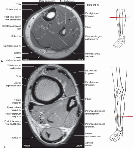
Foot, Ankle, and Calf | Musculoskeletal Key from musculoskeletalkey.com .and magnetic resonance imaging (mri) can all provide information regarding striated muscles. Top suggestions for foot muscle anatomy mri. Posted by radiologyer at 8:12 am. Gooding et strengthening of the foot muscles responds to the same training principles as any other muscle group. This is the first of two parts on the intrinsic muscles of the foot. Musculoskeletal system | muscle structure and function. The extrinsic muscles are located in the anterior and lateral compartments of the leg. Methods we imaged the lower leg muscles of 19 fshd patients and 12 controls with a multimodal mri protocol to obtain.
Subscribe to foot & ankle problems.
Musculoskeletal system | muscle structure and function. Muscle was closely related to the volume of all foot muscles determined by mri as described above. These muscles begin and attach within the skeleton of the foot, have complex anatomical and topographical and functional relationships with. However, on mri images, no muscular abnormalities were detected. Lateral and medial processes of calcaneal tuberosity. Posted by radiologyer at 8:12 am. Mri with hardware in foot? Shop our pre workout and nitric oxide supplements. Indications for foot mri scan. Subscribe to foot & ankle problems. .magnetic resonance imaging (mri) or ultrasound imaging (usi) (soysa et al., 2012; Mri with hardware in foot? In addition, an image of all the muscles of the back and.
Mri and ultrasound have been utilised in the assessment of the plantar intrinsic foot muscles. Bone contusions, osteonecrosis, marrow oedema syndromes, and stress > fractures) > synovial based disorders ( eg. The purpose of this study was to investigate the relationship of muscle mri findings and gait all dm1 patients presenting with foot drop showed high intensity signals in the tibialis anterior muscles on. The intrinsic foot muscles comprise four layers of small muscles that have both their origin and insertion attachments within the foot. Lateral and medial processes of calcaneal tuberosity.
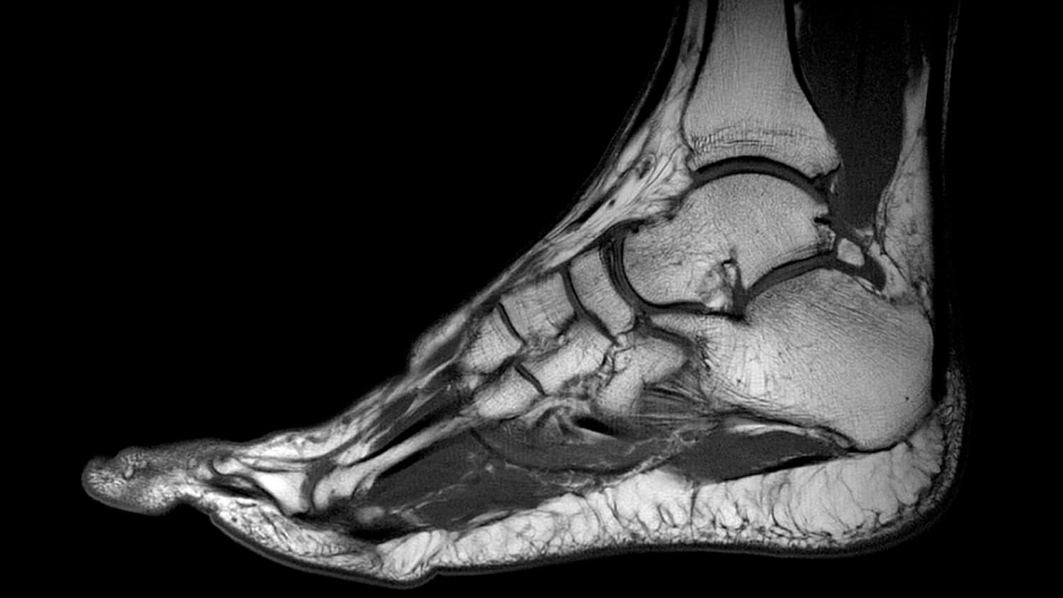
MRI SCAN - Fitter Feet For LifeFitter Feet For Life from fitterfeet.co.uk This article reviews the use of magnetic resonance imaging (mri) in the evaluation of the foot, including a mri of the foot. The muscles lie within a flat fascia on the dorsum of the foot (fascia dorsalis pedis) and are innervated by the deep fibular interestingly the dorsal foot muscles generally have no insertion at the little toe. Lateral and medial processes of calcaneal tuberosity. Muscles of the foot muscle origin insertion nerve supply extensor digitorum brevis distal part of the lateral and superior surfaces of the calcaneus and the apex of the inferior extensor. Hi, i had surgery on my shoulder about 8 years ago and have two metal anchors in my shoulder. Subscribe to foot & ankle problems. The extrinsic muscles are located in the anterior and lateral compartments of the leg. Mri with hardware in foot?
Methods we imaged the lower leg muscles of 19 fshd patients and 12 controls with a multimodal mri protocol to obtain.
By muhammad ali, mb bs; A magnetic resonance imaging (mri) was performed on a normal subject; Foot muscles mri / the extrinsic muscles are located in the anterior and lateral compartments of the leg. Indications for foot mri scan. This is the first of two parts on the intrinsic muscles of the foot. Explore more like foot muscle anatomy mri. Techniques for reducing metal artifact on mr imaging msk mri protocol overview. Posted by radiologyer at 8:12 am. Learn about foot and ankle mri here. Top suggestions for foot muscle anatomy mri. Related posts of foot muscle anatomy mri. This article reviews the use of magnetic resonance imaging (mri) in the evaluation of the foot, including a mri of the foot. Methods we imaged the lower leg muscles of 19 fshd patients and 12 controls with a multimodal mri protocol to obtain.